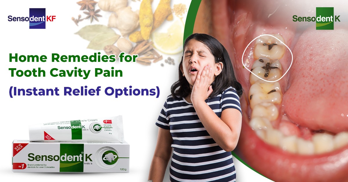

Wisdom tooth cavities are challenging as these third molars erupt late, often in the late teens or early twenties, and are at hard-to-reach spots at the back of the mouth. Decay here leads to intense pain, are at risk of infection, and even cause systemic health issues if ignored. This guide explores symptoms to watch for like underlying causes, effective treatments, and extraction options, so you can make informed decisions about your dental care.
Let’s Understand Wisdom Teeth and Cavities
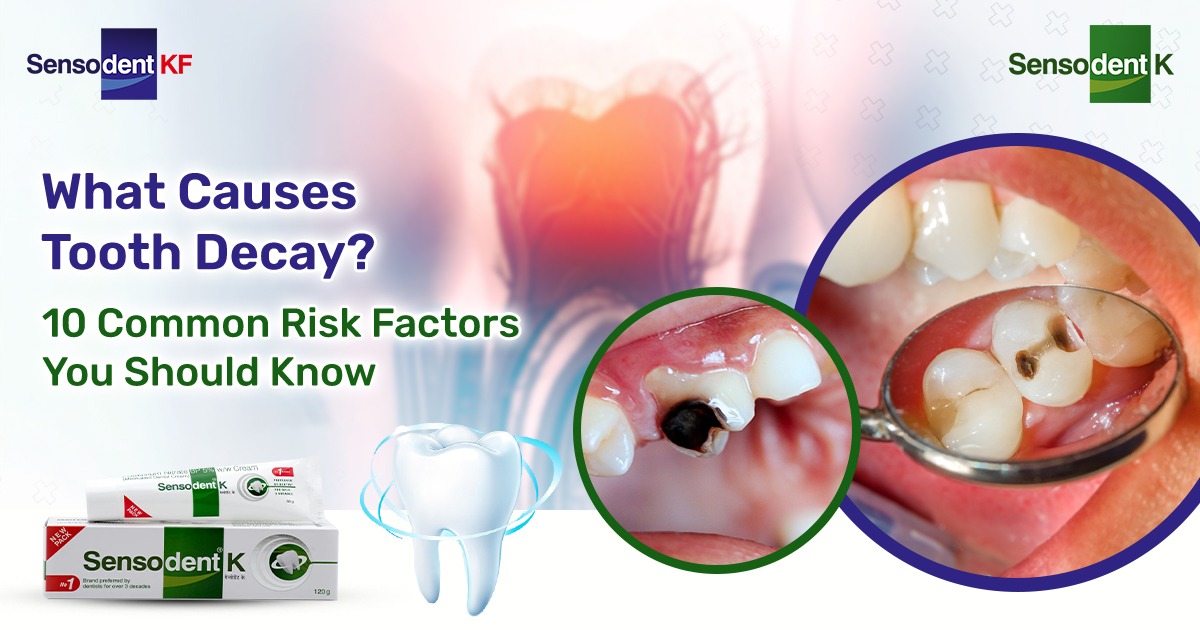
Wisdom teeth, or third molars start emerging between ages 17 and 25, though some people never develop them. Humans generally have four wisdom teeth, two on the top and two on the bottom. Impaction occurs in up to 72% of cases, meaning they don't fully erupt. A cavity, or dental caries, forms when bacteria in plaque produce acids that erode your tooth's enamel, exposing dentin and potentially the pulp.
Cavities in wisdom teeth differ from those in other molars due to limited access for brushing and flossing. Food debris can accumulate there easily, creating an ideal environment for Streptococcus mutans and other bacteria. Without intervention, decay progresses from a white spot lesion to a painful hole with possible abscess formation. Regular dental check-ups with X-rays detect these issues early, as visual inspection alone misses subsurface decay.
Partially erupted wisdom teeth will often trap debris under overlying gum flaps (opercula), fostering pericoronitis alongside cavities. Your genetics can influence tooth size and jaw space. Modern humans have smaller jaws that lead to crowding, exacerbating problems. Maintaining hygiene with tools like water flossers will help, but professional care remains essential.
How to Recognise the Symptoms of Tooth Decay Early
Early detection prevents escalation, yet wisdom tooth cavity symptoms mimic other issues like sinusitis or TMJ disorders. The hallmark is localised pain at the back of the jaw, starting as a dull ache and intensifying to throbbing, especially during chewing hard foods like nuts.
1.Pain Patterns and Sensitivity:
Tooth pain radiates to the ear, temple, or neck due to shared nerve pathways (trigeminal nerve). Sensitivity to hot, cold, or sweet stimuli indicates the breakdown of enamel. It can feel like a sharp zing when sipping coffee or eating chocolate. Nighttime pain worsens as lying down increases blood flow to the head.
2. Visible and Tactile Signs of Tooth Pain:
Look for swelling or redness around the gum line, sometimes forming a gum boil (parulis) draining pus. Bad breath (halitosis) or a metallic taste persists from bacterial overgrowth. Jaw stiffness limits mouth opening (trismus), and swollen lymph nodes under the jaw signal infection spread.
3. Advanced Indicators of Tooth Decay:
Headaches, especially cluster types behind the eye, and facial swelling may be caused by an abscess. Fever, malaise, or difficulty swallowing indicate systemic involvement and it requires urgent care. Impacted teeth may cause adjacent second molar decay, compounding symptoms.
Here’s a quick way to track the symptoms of tooth ache: Note down triggers, duration, and severity. Over-the-counter pain relievers like ibuprofen reduce inflammation temporarily, but consult a dentist promptly.
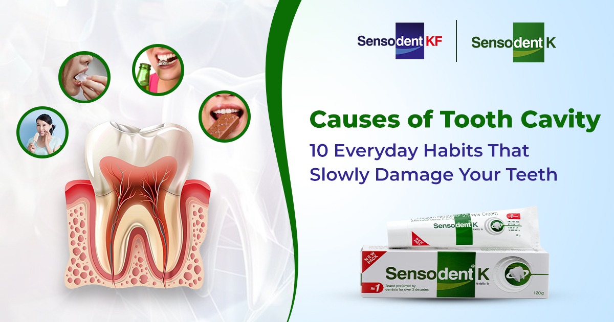
Root Causes of Wisdom Tooth Cavities
A cavity doesn’t appear randomly; there are multiple factors that converge on these vulnerable teeth.
1. Hygiene Challenges:
Brushing reaches only 60-70% of wisdom tooth surfaces effectively. Floss struggles past crowded teeth, allowing plaque to harden into tartar. Tongue scraping misses rear areas, perpetuating the cycle.
2. Anatomical and Tooth Eruption Issues
: Impaction creates stagnant pockets; horizontal or angled eruption abrades cheeks or adjacent teeth. Enamel on wisdom teeth is thinner and more prone to wear from grinding (bruxism). Saliva flow diminishes at night, concentrating acids.
3. Dietary and Lifestyle Contributors:
Eating sugary snacks frequently helps feed bacteria while acidic drinks like sodas erode enamel. Dry mouth from medications (antihistamines, antidepressants), smoking, or dehydration reduces buffering saliva. Infrequent check-ups delay detection.
4. Systemic Risk Factors:
Conditions like diabetes impairs healing and increases decay risk. Poor childhood nutrition affects enamel formation. Genetics play a role in this as some people inherit a softer enamel or crowded arches.
Here are some strategies that help prevent tooth decay include antimicrobial mouthwashes (chlorhexidine), xylitol gums, and electric toothbrushes with small heads. Sealants on partially erupted teeth offer protection, though the application of it may be tricky.
Diagnostic Process of Tooth Decay
A dentist will start with a clinical exam and probe for soft spots while checking bite alignment. Bitewing X-rays reveal interproximal decay; panoramic radiographs assess impaction and root positions. Cone-beam CT scans provide 3D views for complex cases, evaluating bone involvement or nerve proximity.
Transillumination lights will highlight cracks; laser fluorescence devices like DIAGNOdent quantify decay depth numerically. Vitality tests (cold/heat/electric pulp testing) differentiate reversible pulpitis from irreversible. Early diagnosis hinges on patient history—recent pain onset or trauma flags urgency.
| Diagnostic Tool | Purpose | Advantages |
| Bitewing X-ray | Detects proximal decay | Quick, low radiation |
| Panoramic X-ray | Views all wisdom teeth | Comprehensive eruption assessment |
| CBCT Scan | 3D nerve/bone mapping | Ideal for surgical planning |
| Pulp Vitality Test | Assesses nerve health | Differentiates treatable decay |
Non-Extraction Treatments Used by Dentists
Preserving the tooth is ideal when feasible while prioritising function and aesthetics.
1. Fillings and Sealants:
Composite resin fillings bond seamlessly for small cavities, restored in one visit. Glass ionomer cements release fluoride, ideal for high-risk patients. Sealants coat pits/fissures preventively, lasting 5-10 years.
2. Root Canal Therapy (Endodontics):
For pulp-involved decay, access via the crown will remove the infected tissue, clean the canals, and fill it with gutta-percha. Success rates in these procedures exceed 90% in accessible teeth and is followed by a crown for strength. Generally, a challenge will arise with curved roots or incomplete eruption.
When is an Extraction Suggested, and Why
Extraction is the gold standard for untreatable cases. It prevents complications like cyst formation or adjacent tooth damage.
1. Indications for Tooth Removal:
Severe decay can destroy >50% of the tooth structure, can cause recurrent infections, with the resorption of neighbouring roots, or decay risking systemic spread (e.g., endocarditis in heart patients). Prophylactic removal for asymptomatic impactions debates exists; AAOMS guidelines favours early extraction in young adults for easier healing.
2. Simple vs. Surgical Extraction:
Fully erupted teeth undergo simple extraction: local anaesthesia, forceps/elevator, socket cleaning. Surgical extraction for impacted teeth involves flap incision, bone removal (osteotomy), sectioning (coronectomy for nerve risks), and sutures. IV sedation or general anaesthesia suits anxious patients.
3. Advanced Extraction Techniques:
Piezoelectric surgery minimises bone trauma with ultrasonic tips, preserving vitality. Laser-assisted extraction reduces bleeding. For high-risk patients, platelet-rich fibrin (PRF) accelerates socket healing.
| Extraction Type | Procedure Steps | Recovery Time |
| Simple | Anesthesia, loosen, extract | 3-5 days |
| Surgical | Incision, bone removal, sectioning | 7-14 days |
| Coronectomy | Partial tooth removal | Reduces nerve injury |
What is the Recommended Time for Recovery and What is the Aftercare
Post-op swelling peaks at 48 hours, so it is advisable to ice 20 minutes on/off. Consider a soft diet (yogurt, soups) for 1 week and rinse with saltwater after 24 hours. Pain meds (opioids sparingly), antibiotics will be prescribed by the doctor is the area is infected. Avoid straws to prevent dry socket (alveolar osteitis), affecting 2-5%.
Warning signs: excessive bleeding, fever >101°F, or numbness. Full healing takes 3-6 months; bone grafts may precede implants.
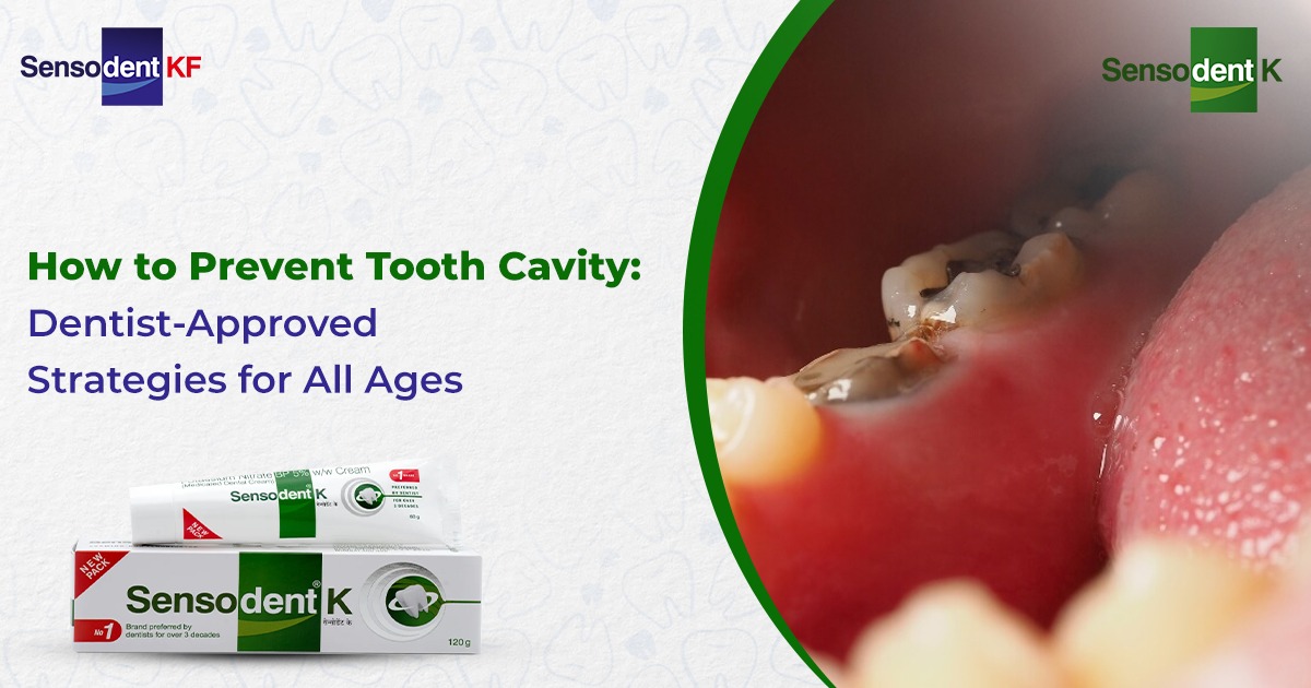
Prevention Strategies
You should brush twice with fluoride toothpaste, flossing wisdom areas, and professional cleanings every 6 months form the base of healthy teeth. Chew sugar-free gum post-meals. Seal high-risk teeth promptly.
Complications and Long-Term Outlook
Untreated cavities risk Ludwig's angina (floor-of-mouth infection) or cavernous sinus thrombosis. This is rare but can be life-threatening. An extraction will lower future decay odds but introduces dry socket or paresthesia (5% inferior alveolar nerve risk).
Most heal uneventfully; space closure occurs naturally in 30%. Orthodontics may realign remaining teeth. Long-term, good habits prevent recurrence elsewhere.
Conclusion: Your Commitment to a Resilient Smile
Wisdom tooth health is an important part of the strong line of defence that protects your overall oral health, just like your tooth enamel does for the rest of your teeth. By understanding the symptoms early, knowing the root causes, and choosing the right treatments or extraction options, you can stay in control of your comfort and your long-term dental health.​
Your smile deserves the best care at every age. Prioritise both your enamel health and your wisdom teeth with SensodentK, so you can enjoy the benefits of a healthy, confident smile for life.